Brain Wave Response to Bottle Color of Herbicides and Non-selective Herbicides in Korea

김 민주  Minju Kim1송 지은  Jieun Song1Sowndhararajan Kandhararjan  Kandhasamy Sowndhararajan1김 성문  Songmun Kim1*

Abstract

The colors of packaging of herbicides and non-selective herbicides on the market in Korea are defined as brown and red, respectively, according to the notification of RDA. The present study aimed to understand consumer’s electroencephalographic (EEG) response when looking at brown and red colors of herbicide and non-selective herbicide packaging papers. The EEG cap was placed on the scalp of each participant (men and women, 10 to 20 years old) and white (control) - brown - white - red colors were sequentially displayed for 5 seconds using the computer monitor. The EEG was measured and statistical analysis was performed using SPSS. For the brown color of the herbicide, men showed a decrease in concentration and a distracting response due to a decrease in the ratio of mid beta to theta (RMT) and the spectral edge of frequency (SEF90). In women, an increase in the ratio of SMR to theta (RSMT) and the spectral edge frequency 50% of the alpha (ASEF) was observed in different brain regions and these EEG changes may enhance the relaxation, stabilization and awakening states of the brain. For the red color of the non-selective herbicide, ASEF increased psychological stability in men. In women, a decrease in absolute high beta (AHB) may associate with a decrease in attention state of the brain. Overall data of the present study clearly revealed that the colors of two herbicides showed significantly different EEG response and gender difference.

Keyword


서 론

우리나라에서 판매되고 있는 농약 품목의 수는 3,007개이며(National Institute of Agricultural Sciences, 2018), 사용목적에 따라 제초제, 생장조정제, 살충제, 살균제, 살비제, 살선충제, 살서제, 식물생장조절제로 구분되고 있다. 농약구분을 위해 농촌진흥청에서는 고시(제2009-2호)를 통해 포장지에 색상을 달리하도록 규정하고 있으며 농약회사에서는 고시에 따라 농약 포장지를 생산하고 있다. 농촌진흥청 고시에 따르면 제초제의 색상은 황색, 비선택성 제초제는 적색, 생장조정제는 청색, 살균제는 분홍색, 살충제는 녹색, 그리고 기타 농약은 백색으로 지정하고 있다.

우리가 색상을 인지하는 과정은 잘 알려져 있다. 눈을 통해 들어온 시각정보는 망막세포, 간상세포와 원추세포를 통과한 후 시각교차를 가로질러 시상에 전달되며, 이어 후두엽에 위치한 일차시각피질에 도달한다. 색상은 일차시각피질에서 대상을 인지하는 경로인 배측 경로(lower ventrical route)를 따라 V4 부위에서 구분되는 것으로 알려져 있다. 그러나 우리가 시각정보를 의식하고, 그 정보에 대해 어떤 방식으로 감정을 가지는지에 대해서는 잘 알려져 있지 않다 (Carter et al., 2009).

색상은 인간의 생리에 영향을 주어 심리 변화뿐만 아니라 행동 변화까지 유도하는데(Elliot and Maier, 2007; Lee et al., 2014; Lee et al., 2017; Mehta and Zhu, 2009) 태양, 불, 피, 위험을 연상시키는 빨간색은 부정적인 사고를 극복하고 활기와 야망을 갖도록 해주고, 새싹, 어린이, 자연을 연상시키는 연두색은 스트레스를 해소시켜주며 마음의 평화를 갖도록 도와주며, 평온, 안식, 생명력을 연상시키는 초록색은 스트레스와 격한 감정을 차분하게 해주고 균형을 잡아주며, 정숙, 차거움, 시원함, 청결을 연상시키는 파랑색은 명상과 휴식을 증가시키고 불면증을 완화시킨다(Kim, 2013). 색상이 인간의 생리에 미치는 영향 때문에 심리학, 마케팅, 감성공학, 건축과 산업디자인 산업에서는 많은 연구가 이루어지고 있다.

본 연구에서 저자들은 국내에서 사용되고 있는 제초제와 비선택성 제초제의 바탕색으로 각각 지정되어 있는 황색과 빨간색이 소비자들에게 미치는 영향을 이해하고자 뇌파(electroencephalogram, EEG)를 측정하고 분석하였다.

재료 및 방법

실험 참가자

2017년 9월 강원대학교 농업생명과학대학 환경융합학부 게시판에 실험 개요를 기재한 설명문을 게시하고 자발적 실험참가자를 모집하였다. 이때 지원한 실험참가자의 연령층은 10-20대이었으며 이들을 대상으로 1:1 면담을 통해 남녀 각 10명씩 총 20명을 선발하였다.

실험 하루 전, 실험참가자들에게 주류, 담배 그리고 약물 섭취를 제한하였으며 피로도가 없도록 충분한 휴식을 취하도록 하였다. 특히 실험 당일엔 실험 방해를 우려하여 정시 식사를 권하였고 1-2시간 전부터는 커피와 흡연을 제한하였다.

뇌파 측정 및 분석

실험참가자를 대상으로 32.4 m2 크기의 실험실에서 최대한 잡음이 혼입되지 않은 안정적인 상태에서 뇌파를 측정하였다. 뇌파측정은 23°C, 습도가 50%가 유지되는 환경에서 실행하였다. 컴퓨터 모니터에 백색(대조군)-황색(제초제)-적색(비선택성 제초제)이 각각 5초씩 나타나도록 프로그램을 하였고, 실험참가자는 편안한 상태에서 모니터를 응시하도록 하면서 3회씩 반복하였다(Fig. 1). 색상은 Microsoft PowerPoint 소프트웨어에서 RGB 값을 지정하였는데 백색은 255,255,255로, 황색은 255,185,9, 그리고 적색은 232,17,35로 설정하였다.

http://dam.zipot.com:8080/sites/wts/images/N0260070205_image/Figure_WTS_7_2_05_F1.jpg

Fig. 1. Schematic drawing of the olfactory system. Redrawn with a permission from the corresponding author Antonio C. Silva Ferreira (Silva Teixeira et al., 2016).

뇌파 측정은 QEEG-64FX 시스템(LAXTHA Inc., Daejeon, Korea)을 사용하여 수행하였고 국제 10-20 시스템에 준용되어 제작된 32 채널 뇌파 캡(EEG cap)을 실험참가자에게 씌우고, reference 전극은 왼쪽 귓볼에 부착하여 측정하였다. 본 연구에서 측정한 뇌파 위치명은 Fp1, Fp2, F3, F4, C3, C4, P3, P4, O1, O2, F7, F8, T7, T8, P7, P8, Afz, Cz, Fz, Pz, Fpz, Oz, Af3, Af4, Fc1, Fc2, Fc5, Fc6, Cp1, Cp2, Cp5, Cp6 이었다.

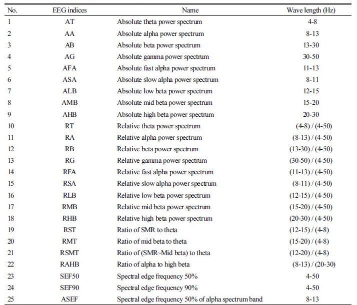
총 3회의 실험 결과 중 잡 피크가 비교적 적은 뇌파 데이터 1개를 선택하여 각각의 색상 마다 시작과 끝의 1초씩을 제외하고 3초간의 결과만을 추출하여 뇌파 분석 프로그램인 Telescan software package (LXSMD61, MAXTHA Inc., Daejeon, Korea)로 데이터를 처리하였다. 데이터 처리에 사용된 지표와 파장은 Table 1에 나타내었다. 데이터는 통계분석 소프트웨어 프로그램인 SPSS (SPSS 18.0 version, SPSS Inc., Chicago, IL, USA)를 이용하여 대응표본 t-검정을 수행하였다.

Table 1. Name and wave length of EEG indices. http://dam.zipot.com:8080/sites/wts/images/N0260070205_image/Table_WTS_7_2_05_T1.jpg

결과 및 고찰

황색 색상에 대한 남성 참가자 반응

우리나라에서 제초제 포장지 색상은 황색이다. 황색은 수용적이고 수동적인 느낌을 주지만 자연과 대지를 상징하는 색으로 대지, 가을, 낙엽, 비옥함, 여유와 같은 상징적 의미를 갖는 것으로 알려져 있다. 황색은 노랑과 빨강 사이에 있는 색으로 색감이 풍부하고 심오한 색으로 생산력, 모성적인 힘 그리고 자연과의 일치를 의미한다. 뿐만 아니라 활력과 생명력의 감소, 불모의 대지를 연상시키기도 한다. 컬러테라피에서 황색은 신경전달물질 세로토닌의 합성 촉진, 프로스타글란딘 E1 형성 촉진으로 만성 피로감을 약화시켜 강박증이나 노이로제가 있는 환자들에게 사용되고 있다(Kim, 2013).

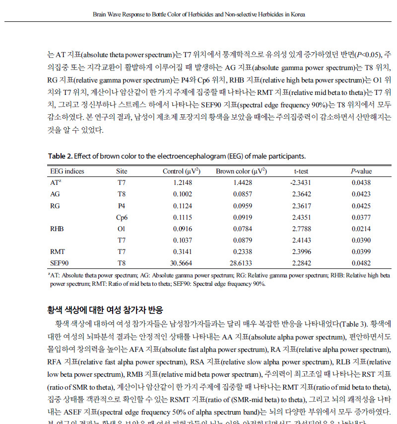
본 연구에서 제초제 포장지의 황색 색상(RGB값 255:127:0)에 대한 소비자들의 반응을 이해하기 위하여 뇌파를 측정한 결과(Table 2), 25개의 뇌파지표 중 졸리거나, 얕은 수면상태에 있거나, 혹은 깊은 명상을 할 때 주로 나타나는 AT 지표(absolute theta power spectrum)는 T7 위치에서 통계학적으로 유의성 있게 증가하였던 반면(P<0.05), 주의집중 또는 지각교환이 활발하게 이루어질 때 발생하는 AG 지표(absolute gamma power spectrum)는 T8 위치, RG 지표(relative gamma power spectrum)는 P4와 Cp6 위치, RHB 지표(relative high beta power spectrum)는 O1 위치와 T7 위치, 계산이나 암산같이 한 가지 주제에 집중할 때 나타나는 RMT 지표(relative mid beta to theta)는 T7 위치, 그리고 정신부하나 스트레스 하에서 나타나는 SEF90 지표(spectral edge frequency 90%)는 T8 위치에서 모두 감소하였다. 본 연구의 결과, 남성이 제초제 포장지의 황색을 보았을 때에는 주의집중력이 감소하면서 산만해지는 것을 알 수 있었다.

Table 2. Effect of brown color to the electroencephalogram (EEG) of male participants. http://dam.zipot.com:8080/sites/wts/images/N0260070205_image/Table_WTS_7_2_05_T2.jpg

zAT: Absolute theta power spectrum; AG: Absolute gamma power spectrum; RG: Relative gamma power spectrum; RHB: Relative high beta power spectrum; RMT: Ratio of mid beta to theta; SEF90: Spectral edge frequency 90%.

황색 색상에 대한 여성 참가자 반응

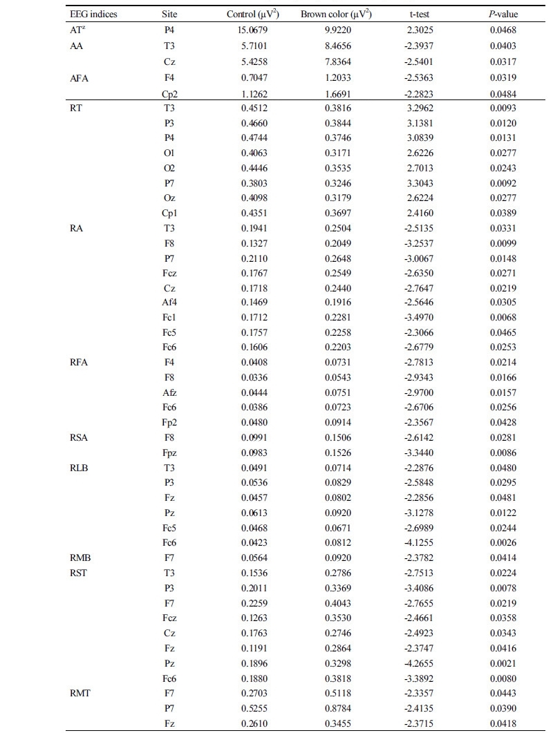
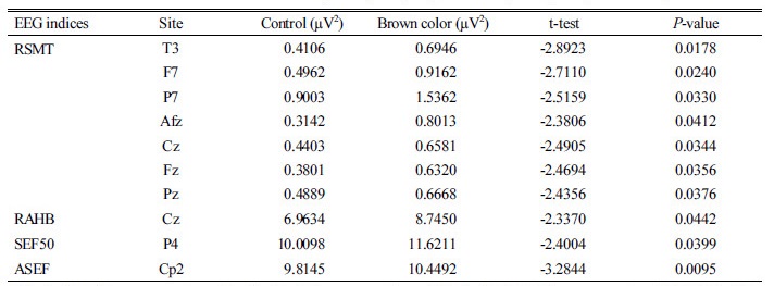
황색 색상에 대하여 여성 참가자들은 남성참가자들과는 달리 매우 복잡한 반응을 나타내었다(Table 3). 황색에 대한 여성의 뇌파분석 결과는 안정적인 상태를 나타내는 AA 지표(absolute alpha power spectrum), 편안하면서도 몰입하여 창의력을 높이는 AFA 지표(absolute fast alpha power spectrum), RA 지표(relative alpha power spectrum), RFA 지표(relative fast alpha power spectrum), RSA 지표(relative slow alpha power spectrum), RLB 지표(relative low beta power spectrum), RMB 지표(relative mid beta power spectrum), 주의력이 최고조일 때 나타나는 RST 지표(ratio of SMR to theta), 계산이나 암산같이 한 가지 주제에 집중할 때 나타나는 RMT 지표(ratio of mid beta to theta), 집중 상태를 객관적으로 확인할 수 있는 RSMT 지표(ratio of (SMR-mid beta) to theta), 그리고 뇌의 쾌적성을 나타내는 ASEF 지표(spectral edge frequency 50% of alpha spectrum band)는 뇌의 다양한 부위에서 모두 증가하였다. 본 연구의 결과는 황색을 보았을 때 여성 피험자들의 뇌는 이완, 안정화되면서도 각성되었음을 나타낸다.

Table 3. Effect of brown color to the electroencephalogram (EEG) of female participants. http://dam.zipot.com:8080/sites/wts/images/N0260070205_image/Table_WTS_7_2_05_T3.jpg
Table 3. Effect of brown color to the electroencephalogram (EEG) of female participants (Continued). http://dam.zipot.com:8080/sites/wts/images/N0260070205_image/Table_WTS_7_2_05_T3_1.jpg

zAT: Absolute theta power spectrum; AA: Absolute alpha power spectrum; AFA: Absolute fast alpha power spectrum; RT: Relative theta power spectrum; RA: Relative alpha power spectrum; RFA: Relative fast alpha power spectrum; RSA: Relative slow alpha power spectrum; RLA relative low beta power spectrum; RMB: Relative mid beta power spectrum; RSMT: Ratio of (SMR - mid beta) to theta; RAHB: Ratio of alpha to high beta; SEF50: Spectral edge frequency 50%; ASEF: Spectral edge frequency 50% of alpha spectrum band.

적색 색상에 대한 남성 참가자 반응

우리나라 제초제 중 비선택성 제초제 포장지 색상은 적색이다. 일반적으로 적색은 시대와 문화적 차이에도 불구하고 열정, 생명, 환희, 분노, 공격성, 흥분, 위험, 유혹, 에로스, 역동적, 적극적인 의미를 나타낸다. 적색의 연상 대상은 태양, 피, 불꽃, 공산국가, 네온, 장미, 우체통, 딸기, 소방차, 셀비어, 오똑이, 투우사, 화재, 스포츠카, 사과, 립스틱, 토마토이며, 심리적으로는 부정적인 사고를 극복할 수 있도록 하며, 활기와 야망을 갖게 하는 것으로 알려져 있다. 컬러테라피에서 적색은 혈압을 높이고 아드레날린을 분비시켜 활동 향상에 도움을 주기 때문에 소심증이나 우울증 치료, 또는 무감각하고 냉정하며 너무 생각이 많은 사람들에게 추천하여 내면적인 주의집중을 분산시켜 밖으로 쏠리게 하여 치료에 활용하고 있다(Kim, 2013).

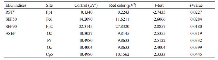
비선택성 제초제 포장지의 적색 색상에 대한 남성들의 뇌활성 반응을 알기 위해서 적색(RGB 값 255:0:0) 색상을 보여주면서 뇌파를 측정한 결과, 절대 뇌파 스펙트럼 중 통계학적으로 유의성 있는 차이가 나는 지표는 없었다(Table 4). 그러나 적색 색상을 본 남성들은 RST 지표와 SEF90 지표가 증가하였는데 RST 지표는 주의력이 최고조일 때 발생하는 것으로 알려져 있다. 그리고 쾌적성을 나타내는 ASEF 지표(spectral edge frequency 50% of alpha spectrum band)와 SEF50 지표는 감소되었는데 이와 같은 결과들은 적색 색상에 대해 남성들은 주의력이 최고조인 상태에서 긴장을 하였다는 것을 의미한다.

Table 4. Effect of red color representing non-selective herbicides to the electroencephalogram (EEG) of male participants. http://dam.zipot.com:8080/sites/wts/images/N0260070205_image/Table_WTS_7_2_05_T4.jpg

zRST: Ratio of SMR to theta; SEF50: Spectral edge frequency 50%; SEF90: Spectral edge frequency 90%; ASEF: Spectral edge frequency 50% of alpha spectrum band.

적색 색상에 대한 여성 참가자 반응

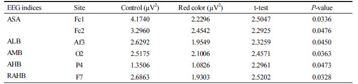
여성들에게 적색을 보여주었을 때, 여성들은 이완이나 휴식하면서도 언제든지 활동할 수 있게 집중하고 있는 상태를 나타내는 ASA 지표(absolute slow alpha power spectrum), 주의나 경계할 때 나타나는 ALB 지표(absoltue low power spectrum), 집중과 주의를 나타내는 AMB 지표(absoltue mid beta power specturm), 스트레스, 정신부하, 또는 긴장 시에 나타난 AHB 지표(absolute high beta power spectrum) 뿐만 아니라 안정 및 이완도를 나타내는 RAHB 지표(ratio of alpha to high beta)는 모두 감소하였다(Table 5). 본 연구의 결과는 적색 색상에 대해서 여성들은 집중, 주의, 경계력을 낮추고 산만함을 유도하였다는 것을 알 수 있다.

Table 5. Effect of red color representing non-selective herbicides to the electroencephalogram (EEG) of female participants. http://dam.zipot.com:8080/sites/wts/images/N0260070205_image/Table_WTS_7_2_05_T5.jpg

zASA: Absolute slow alpha power spectrum; ALB: Absolute low beta power spectrum; AMB: Absolute mid beta power spectrum; AHB: Absolute high beta power spectrum; RAHB: Ratio of alpha to high beta.

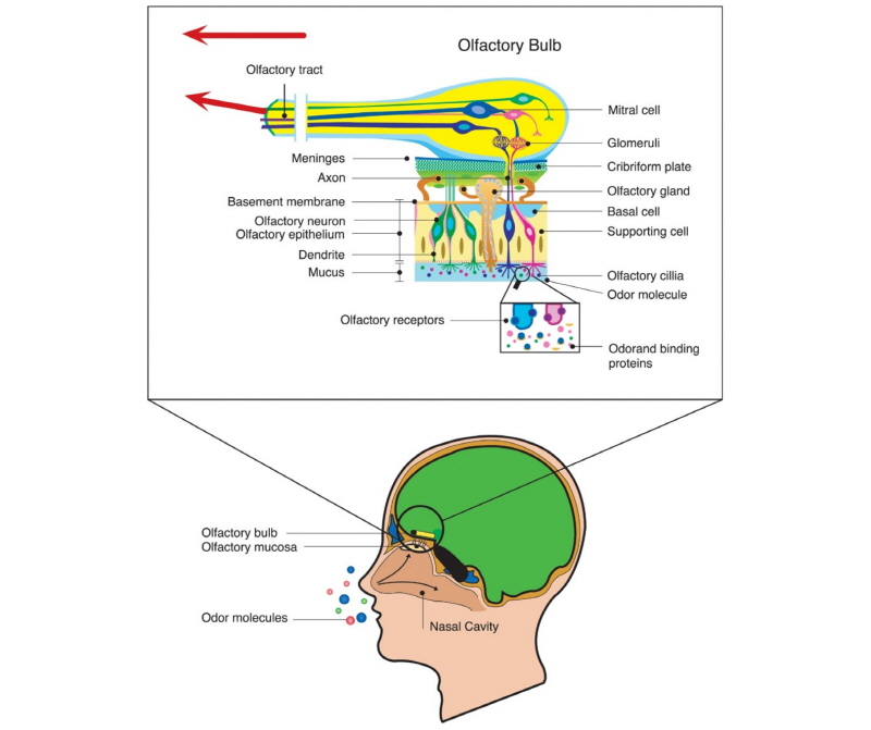
남성과 여성의 뇌는 해부학적으로 차이가 있는 것으로 많은 연구자들은 보고하고 있다(Coggrove et al. 2007). 남성은 여성과 비교하여 시상하부(hypothalamus)가 더 크고, 뇌량(corpus callosum)은 더 작은 것으로 알려져 있다(Zaidi, 2010). 그리고 여성은 후각망울(olfactory bulb)에 신경(neuron)과 신경교세포(glial cell)를 더 많이 가지고 있으며(Oliveira-Pinto et al., 2014), 사회적 인식과 기술과 관련이 있는 배측 전두엽 피질(ventral frontal cortex)이 더 발달해 있고, 사회적 유대와 관련이 있는 직선이랑(straight gyrus)이 남성과 비교해서 약 10% 더 컸다(Wood et al., 2008).

그리고 남성과 여성의 뇌는 기능적으로도 차이가 있는 것으로 알려져 있다. Tunҫ et al. (2016)은 남성의 경우 운동(motor), 감각(sensory) 및 관리기능(executive function)에서 커넥텀이 더 발달해 있고, 여성의 경우에는 사회관계, 주의력과 기억과제에서 커넥텀이 더 발달해 있으며, 휴식기의 뇌 활동을 EEG로 측정할 때 여성은 남성과 비교하여 두정-후두엽 부위(parieto-occipital area)에서 β파와 γ파를 더 많이 내는 것으로 나타났다(Jausovec and Jausovec, 2010). 이러한 차이에 대해서 Zaidi (2010)는 지각, 인지, 기억, 신경기능에 있어서 남성과 여성간의 차이는 유전, 호르몬, 환경 요인에 의해 복합적으로 나타나는 것이라 하였다.

본 연구에서 남성과 여성에게 동일한 황색(Table 2, 3)과 적색(Table 4, 5) 색상을 보여주었을 때 뇌 반응은 매우 달랐다. 색상에 대한 남녀 성별 차이에 대해서는 여러 연구자들이 본 연구에서의 결과와 유사한 결과를 보고한 바 있다. 그리고 남녀 간에는 색상뿐만 아니라 향에 대한 반응도 다르다고 보고되었다(Kim et al., 2017; Sowndhararajan and Kim, 2016). Park (2013)은 남자/여자 어린아이들이 선호하는 색상이 별도로 있으며 성별 간 차이가 크다는 것을 보고하였다. 남자 아이들과 비교하여 여자 어린아이는 적색과 보라 색상을 더 좋아하였고, 적색에 대해서도 남자 아이들은 명도가 낮은 빨간 색상을 선호하였던 반면 여자 아이들은 명도가 높은 빨간 색상을 좋아하였다. 그리고 Radeloff (1990)는 매력을 나타내는 색상에 대한 연구에서 남성은 여성보다 밝은 색상을 더 선호하지만 부드러운 색에 대해서는 선호도가 낮다는 것을 보고하였다.

색상-뇌파 연구에서는 색상을 보여주면서 EEG를 측정하였는데 Trikha (2010)는 남성과 여성에게 2개의 색상을 보여주면서 사건관련전위(event-related potentials, ERPs)를 측정하였을 때 남성이 여성보다 P300에서 높은 ERPs 값을 보였다는 것을 보고하였고, Wang and Shih (2017)는 60명의 젊은 남성과 여성을 대상으로 semi-figurative 이미지 또는 추상적 이미지의 색상을 보여주면서 EEG를 측정한 결과 남녀 간의 색상 차이는 없었다고 보고하였는데, 이러한 결과는 색상만을 보여주면서 EEG를 측정한 본 연구의 결과와는 달랐다. Wang and Shih (2017)의 연구에서는 이미지와 색상이 동시에 실험참가자에게 제시되면서 EEG가 측정되었던 반면, 본 연구에서는 단지 색상만이 제시되면서 EEG가 측정되었기 때문에 상이한 결과가 도출되었다고 판단된다.

References

1 Carter, R., Aldridge, S., Page, M. and Parker, S. 2009. The Human Brain Book. 21 Century Books, Paju, Korea. (In Korean) 

2 Coggrove, J.P., Mazure, C.M. and Staley, J.K. 2007. Evolving knowledge of sex differences in brain structure, function and chemistry. Biol. Psychiatry 62(8):847-855.  

3 Elliot, A.J. and Maier, M.A. 2007. Color and psychological functioning. Curr. Dir. Psychol. Sci. 16(5):250-254. 

4 Jausovec, N. and Jausovec, K. 2010. Resting brain activity: Differences between genders. Neuropsychologia 48:3918-3925.  

5 Kim, M., Kandasamy, S., Kim, T., Kim, J.E., Yang, J.E., et al. 2017. Gender differences in electroencephalographic activity in response to the earthy odorants geosmin and 2-mentylisoborneol. Applied Sci. 7:876 doi:10.3390/ app7090876. 

6 Kim, S.H. 2013. Color Psychology. Idam Book, Paju, Korea. (In Korea) 

7 Lee, H., Kim, S., Kim, K. and Hong, K. 2017. Brain waves evoked by the changes of background pastel colors with a pattern of achromatic color. Fashion & Text. Res. J. 19(5):653-660.  

8 Lee, H.W., Kim, H.N., Kim, Y.C., Jeong, Y.S. and Lee, J.S. 2014. The brain wave analysis of the color stimulations in red, green and blue according to the gender and age. Korea Society of Color Studies 2014 Fall Conference 37-41. (In Korean)  

9 Mehta, R. and Zhu, R.J. 2009. Blue or Red? Exploring the effect of color on cognitive task performances. Science 323:1226-1229.  

10 National Institute of Agricultural Sciences. 2018. http://pis.rda.go.kr/registstus/agchmRegistStus/prdlstInqire.do (Accessed Apr. 11, 2018) 

11 Oliveira-Pinto, A.V., Santos, R.M., Coutinho, R.A., Oliveira, L.M., Santos, G.B. et al. 2014. Sexual dimorphism in the human olfactory bulb: Females have more neurons and glial cells than males. PLOS ONE 9(11):1-9. 

12 Park, J.G. 2013. Correlations between color attributes and children’s color preferences. Color Res. Appl. 39(5): 452-462.  

13 Radeloff, D.J. 1990. Role of color in perception of attractiveness. Perceptual and Motor Skilss. 71(1):151-160. 

14 Silva Teixeira, C.S., Cerqueira, N.M.F.S.A., Silva Teixeira, A.C. 2015. Unravelling the olfactory sense: From the gene to odor perception. Chem. Senses 41:105-121.  

15 Sowndhararajan, K. and Kim, S. 2016. Influence of fragrances on human psychophysiological activity: With special reference to human electroencephalographic response. Sci. Pharm. 84(4):724-751.  

16 Trikha, A. 2010. Gender and color specific differences in event related potentials. MS Thesis, Brigham Young Univ., Provo, Utah, USA. 

17 Tunҫ, B., Solmaz, B., Parker, D., Satterthwaite, T.D., Elliott, M.A., et al. 2016. Establishing a link between sex-related differences in the structural connectome and behaviour. Phil. Trans. R. Soc. B 371:20150111.  

18 Wang, C.H. and Shih, Y.H. 2017. Investigating the impact of coloring experience on young adults through brainwave variations and image preference. US-China Education Review B. 7(10):449-458. 

19 Wood, J.L, Heitmiller, D., Andreasen, N.C. and Nopoulos, P. 2008. Morphology of the ventral frontal cortex: Relationship to femininity and social cognition. Cereb Cortex 18:534-540.  

20 Zaidi, Z.F. 2010. Gender differences in human brain: A review. The Open Anatomy J. 2:37-55.