First Report of Red Thread of Kentucky Bluegrass (Poa pratensis L.) Caused by Laetisaria fuciformis on Golf Course in Koreaz

The Korean Society of Weed Science and The Turfgrass Society of Korea
이 정한  Jung Han Lee1김 정호  Jeong Ho Kim1심 규열  Gyu Yul Shim1곽 연식  Youn-Sig Kwak2

Abstract

In the middle of May 2018, typical red thread disease symptoms were observed on Kentucky bluegrass (Poa pratensis L.) on a golf course, which locates at Yangsan, Gyeongnam province in Korea. Irregular-shaped patched symptoms were observed in both fairway and rough of golf course. The foliar symptom was dried out and faded to straw color and tip of the grass leaves were tangled like thread. Early morning, infected and tangled leaves were covered with the pinkish gelatinous antler-like structure (sclerotinia) as a typical red thread disease symptom. Causal fungal pathogens were isolated from the symptom in Kentucky bluegrass, Cultural characteristic on potato dextrose agar, color of colony was pale pink and conjugated hyphae, sclerotium of irregular shape was pale pink and 3-5 mm diameter in size. The pathogen was identified as Laetisaria fuciformis based on morphological and culture characteristics as well as molecular characteristics. Pathogenicity test was verified on the Kentucky bluegrass by Koch’s postulates. This is the first report of red thread disease occurrence in Kentucky bluegrass by L. fuciformis in Korea.

Keyword


서 론

켄터키블루그래스(Poa pratensis L.)는 한지형 잔디로서 북방지역이나 전이지대에서 다양한 목적으로 사용되며 미국에서는 골프장이나 축구장과 같이 집약적인 관리가 요구되는 고품질 스포츠 경기장에 사용된다(Meyer and Funk 1989; Porceddu et al., 2002). 국내 골프장에서도 티잉그라운드, 그린칼라, 페어웨이 등에 사용된다(Lee, 2012). Red thread의 원인균인 Laetisaria fuciformis의 기주는 벤트그래스(Agrostis stolonifera), 훼스큐류(Festuca spp.), 페레니얼라이그래스(Lolium perenne L.), 에뉴얼블루그래스(Poa annua) 및 켄터키블루그래스로 알려져 있으며(Johnson and Bowyer, 1982) 오스트레일리아, 캐나다. 미국, 영국, 네덜란드와 아일랜드 등에서 발생이 보고되어 있다(Kaplan and Jackson, 1983; McAlpine, 1906; Stalpers and Loerakker, 1982; Wakefield, 1917). 하지만 국내에서는 Red thread에 대한 연구 보고가 없는 상태이다. 본 연구에서는 경남 양산에 소재하고 있는 에덴밸리골프장의 켄터키블루그래스 병반으로부터 병원균을 분리, 동정하여 L. fuciformis에 의한 red thread의 국내 발생을 처음으로 보고하고자 한다.

발병 및 병징

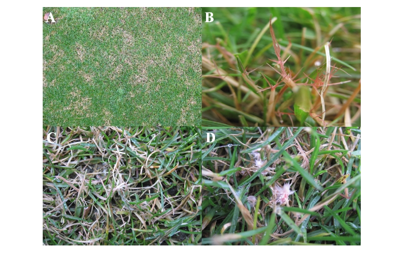
2018년 5월 중순, 경남 양산에 소재하고 있는 에덴밸리골프장의 켄터키블루그래스 페어웨이와 러프에 부정형의 붉은 빛을 띄는 병반이 발생하였다. 병반은 직경 10-50 cm 정도의 크기로 형성되었으며 병반 부위의 잔디 잎은 볏짚색으로 마름증상을 보이며 고사하였다(Fig 1A). 감염된 잎의 끝 부분에는 길이 0.5-10 cm 크기의 사슴뿔 모양의 젤라틴화된 균핵(sclerotia)이 형성되었다 (Fig. 1B). 병반 부위의 잎의 끝부분은 실모양으로 헝클어져 있었으며(Fig. 1C), 상대습도가 높으면 기중균사가 형성되어 뭉쳐져 분홍색의 솜털 모양을 형성하였다(Fig. 1D). 병은 초기에 감염된 잎이 수침상으로 변하고 녹색이 점점 옅어지고 마르면서 황갈색으로 고사하였으며, 이후 병세가 진전되면서 불규칙한 병반을 형성하였고, 서로 결합하여 더 큰 병반을 형성하였다.

http://dam.zipot.com:8080/sites/wts/images/N0260070311_image/Figure_WTS_7_3_11_F1.jpg

Fig. 1. Symptoms and signs of red thread disease in Kentucky bluegrass. A: Red thread on golf course growing Kentucky bluegrass; B: Antler like structures (sclerotia) growth from blighted leaves; C: connecting leaf blades; D: flocks of pink mycelium on leaf blades.

L. fuciformis는 0-30oC에서 생장할 수 있으며 건조한 상태에서 2년까지 견딜 수 있다. 발생조건은 16-24oC 정도의 서늘하고 습한 조건에서 발생하는 것으로 알려져 있다(Smiley et al., 2005). 본 연구에서 red thread 발생 지역은 해발 700-800 m로 고도가 높으며 이 때문에 5월 중순에 병발생이 시작되어 6월 말까지도 병반이 관찰되었다.

병원균 분리 및 동정

병원균 분리를 위하여 10 cm 크기의 병반에서 감염된 잎을 채취하였다. 감염된 잎을 0.5 cm2으로 자르고 NaOCl (1%) 용액에서 30초간 표면 살균하였으며 에탄올(70%) 용액에 다시 30초간 살균한 후 멸균수로 3회 세척하고 수분 제거 후 water agar (WA; agar 20g/L)에 치상하여 20oC 항온기에서 배양하였다. 배양 3일 후 시료 조직에서 자란 균사를 potato dextrose agar (PDA; potato dextrose 15 g, agar 20 g/L)로 옮겨 배양하였다.

http://dam.zipot.com:8080/sites/wts/images/N0260070311_image/Figure_WTS_7_3_11_F2.jpg

Fig. 2. Mycological characteristics of Laetisaria fuciformis isolated from Kentucky bluegrass. A: Colony, and sclerotinia which the red arrow indicates on potato dextrose agar (PDA) grown at 20°C in 7 days; B: Probasidium; C & D: Hyphae and strands of conjugated hyphae.

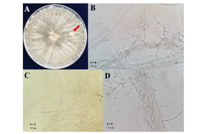
PDA에서 2주일간 배양한 균총은 연한 분홍색을 띄며 젤라틴 형태의 균사다발이 팬(fan) 모양으로 휘어져 자라며 균사다발을 중심으로 균사가 빗자루 모양으로 자라는 것이 관찰되었다. 또한 PDA배지내에서 균핵이 형성되었고(Fig. 2A). 12-20×5-10 um 크기의 불규칙한 모양인 전담자기(probasidia)가 형성되는 것을 관찰하였다(Fig. 2B) PDA에서 배양된 병원균 균사는 직경이 3-7 um이고 격막(septum)이 있으며 다핵성세포(polynuclear cell)였다(Fig. 2C). 균사가 다발형태로 뭉쳐져 있는 균사다발이 빈번하게 관찰되었다(Fig. 2D). 이는 Stalpers and Loerakker (1982)가 보고한 L. fuciformis의 배양 특징과 유사하였다.

분리된 병원균의 계통학적 분석을 위해 potato dextrose broth (PDB)에 5일간 액체 배양한 후 균사체를 멸균수에 세척하였다. 균사체에서 CTAB 추출방법(Graham et al., 2003)을 이용하여 total genomic DNA를 추출하였으며 ITS1F/ITS4 primer set를 이용하여 internal transcribed spacer (ITS) 영역의 유전자 염기서열을 증폭하였다(White et al., 1990). Polymerase chain reaction (PCR) 반응조건은 94°C에서 5분간 pre-denaturation을 진행한 후 94°C에서 30초간 denaturation, 55°C에서 45초간 annealing, 72°C에서 40초간 extension을 수행하였으며 이를 총 30 cycle 수행 후 마지막으로 72°C에서 10분간 final extension을 수행하였다. 증폭된 PCR 산물을 1% agarose gel에서 전기영동을 한 후 정제하였으며 염기서열 분석을 수행하였다. ITS 영역의 염기서열분석은 autosequencer (ABI 3030, Applied Biosystems, Foster City, CA, USA)로 수행하였으며 확보된 염기서열은 GenBank database (National Centre for Biotechnology Information) 데이터베이스의 NCBI BLAST 검색프로그램 (https://blast.ncbi.nlm.nih.gov/Blast.cgi)을 이용하여 Laetisaria 속 근연종의 염기서열을 수집하였다. 계통분석은 MEGA 7.0 프로그램() (Kumar et al., 2016)에서 ClustalW법으로 정렬하고 neighbor-joining (NJ) 방법으로 계통도를 작성하였다(Saitou and Nei, 1987). Sequence distance는 Tajima-Nei parameter model로 계산하였으며(Tamura et al., 2007) outgroup으로 Botrytis cinerea (GenBank accession No. KU992699.1)를 사용하였다. 그 결과 켄터키블루그래스에 red thread를 일으키는 균주의 ITS 영역의 크기는 701 bp였으며, NCBI database에 등록된 ITS sequence 중 AY613910, AY613913, AY613912, AY613909와 99-100%로 일치하였다(Fig. 3). 따라서 병징에서 분리된 병원균은 형태학적, 유전학적으로 L. fuciformis로 동정되었다.

http://dam.zipot.com:8080/sites/wts/images/N0260070311_image/Figure_WTS_7_3_11_F3.jpg

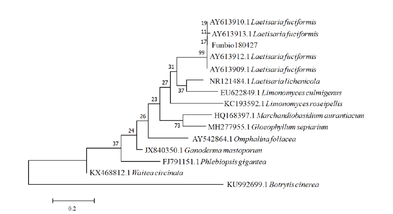
Fig. 3. Phylogenetic identification of Laetisaria fuciformis based on internal transcribed spacer (ITS) rRNA sequences. Numerical values on branches are bootstrap values calculated as a percentage of bootstrap replications from 1000 replication. L. fuciformis Funbio180427 represented the isolate. A phylogenetic tree inference was conducted with the MEGA 7.0 program and phylogenetic distances were calculated using the neighbor-joining method. Bar=0.2 distance between samples. Botrytis cinerea (GenBank accession No. KU992699.1) served as out-group references (Weir et al. 2012).

http://dam.zipot.com:8080/sites/wts/images/N0260070311_image/Figure_WTS_7_3_11_F4.jpg

Fig. 4. Pathogenicity test of Laetisaria fuciformis in the Kentucky bluegrass. A: symptoms at 4 days after inoculation; B: Symptoms on individual leaves.

병원성 검정

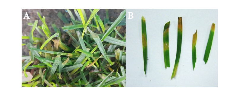
감염된 잎에서 분리된 L. fuciformis를 PDA배지에서 2주간 배양하여 병원성 검증에 이용하였다. 병원균 균사체가 포함된 배지를 메스로 5 mm2로 절단하고 한천 조각을 켄터키블루그래스(Midnight)의 잎에 치상하였으며 대조구로는 병원균이 없는 WA를 치상하였다. 접종된 잔디는 20±1℃, 상대습도 90%가 유지되는 생장상(Table Top B.O.D Incubator, DASOL SCIENTIFIC, Hwaseong, Korea)에서 병징이 발현될 때까지 유지하였다. 병징 발현 후 앞서 언급한 병원균 분리방법을 이용하여 병원균을 재분리하였으며 PDA에 배양하여 균총모양, 균사 및 전담자기 등의 균학적 특성을 비교, 확인하였다. 병원성 검정 결과, 병원균이 접종 3일 후부터 잔디 잎 부분이 수침상으로 변하였으며, 4일 후에는 황화되는 것을 확인하였다(Fig. 4). 황화된 부위로부터 병원균을 재분리하여 PDA에 배양한 결과, 접종한 원래의 균과 같은 균학적 배양 특성을 나타냄을 확인할 수 있었다.

이상과 같이 골프장의 켄터키블루그래스에 붉은 색의 마름현상을 보이는 병징으로 부터 병원균을 분리, 동정하였고, 분리한 균의 접종시험을 통하여 병원성을 확인한 바, Laetisaria fuciformis에 의한 red thread로 동정하였다. 본 논문은 red thread에 관한 한국에서의 첫 보고이므로 저자들은 red thread의 한국명을 ‘붉은뿔마름병’으로 제안한다.

References

1  Graham, J., Marshall, B. and Squire, G. 2003. Genetic differentiation over a spatial environmental gradient in wild Rubus idaeus populations. New Phytol. 157:667-675.  

2  Johnson, B. J. and Bowyer, T. H. 1982. Management of herbicide and fertility levels on weeds and Kentucky bluegrass turf. Agron. Journal. 74:845-850.  

3  Kaplan, J.D. and Jackson, N. 1983. Red thread and pink patch disease of turf grasses. Plant Dis. 67:159-162.  

4  Kumar, S., Tamura, K. and Tamura, K. 2016. MEGA7: Molecular evolutionary genetics analysis version 7.0 for bigger datasets. Mol. Biol. Evol. 33(7):1870–1874.  

5  Lee, S.K. 2012. Irrigation frequency for Kentucky Bluegrass (Poa pratensis) growth. Asian J. Turfgrass Sci. 26(2):123-128.  

6  McAlpine, D. 1906. A new hymenomycete - the so called Isaria fuciformis. Annal. Mycol. 4:541-551.  

7  Meyer, W.A. and Funk, C.R. 1989. Progress and benefits to humanity from breeding cool-season grasses for turf. pp. 31-48. In: Sleeper, D.A., Asay, K.H. and Pedersen, J.F. (Eds.) Contributions from breeding forage and turf grasses. Crop Science Society of America Special Publication, Madison, WI, USA.  

8  Porceddu, A., Albertini, E., Barcaccia, G., Falistocco, E. and Falcinelli, M. 2002. Linkage mapping in apomictic and sexual Kentucky bluegrass (Poa pratensis L.) genotypes using a two way pseudo-testcross strategy based on AFLP and SAMPL markers. Theor. Appl. Genet. 104:273-280.  

9  Saitou, N. and Nei, M. 1987. The neighbor-joining method: a new method for reconstructing phylogenetic trees. Mol. Biol. Evol. 4:406-25.  

10  Smiley, R.W., Dernoeden, P.H. and Clarke, B.B. 2005. Compendium of turfgrass diseases, 3rd ed. APS press, MN, USA.  

11  Stalpers, J.A. and Loerakker, W.M. 1982. Laetisaria and Limonomyces species (Corticiaceae) causing pink diseases in turf grasses. Can. J. Bot. 60(5):529-537.  

12  Tamura, K., Dudley, J., Nei, M. and Kumar, S. 2007. MEGA4: molecular evolutionary genetics analysis (MEGA) software version 4.0. Mol. Biol. Evol. 24:1596-1599.  

13  Wakefield, E.M. 1917. Notes on British thelephoraceae. Trans. Br. Mycol. Soc. 5:474-481.  

14  Weir, B.S., Johnston, P.R. and Damm, U. 2012. Colletotrichum gloeosporioides species complex. Stud. Mycol. 73:115-180.  

15  White, T.J., Bruns, T.D., Lee, S.B. and Taylor, J.W. 1990. Amplification and direct sequencing of fungal ribosomal RNA genes for phylogenetics pp. 315-322. In: Innis, M.A., Gelfand,D.H. and Sninsky, J.J, (Eds.). PCR protocols: a guide to methods and applications. N.Y: Academic Press, NY, USA.