서 언
한국잔디(Zoysiagrass)는 난지형 잔디로 균일한 질감을 가지며 내마모성이 우수하고 한지형 잔디와 비교하여 내병성이 높으며 국내 골프장에서는 페어웨이나 티잉그라운드용으로 사용된다(Beard, 1973; Geon et al., 2005). 국내에서 한국잔디에 발생하는 병으로는 라이족토니아마름병(large patch, Rhizoctonia solani AG2-2 [IV]), 잔디 녹병(rust, Puccinia Zoysiae), 잔디 모잘록병(blight, Curvularia geniculata), 동전마름병(dollar spot, Sclerotinia homoeocarpa), 테이크올패취(take-all patch, Gaeumannomyces graminis), 잔디 노균병(downy mildew, Scleropthora macrospora), 웨이티아링패취(Waitea ring patch, Waitea circinata), 봄마름병(spring dead spot, Rhizoctonia cerealis)이 보고되어있다(Cha et al., 2012; Cheon et al., 2016; Kim and Lee, 1973; Kim et al., 1991; Kim et al., 2014; Shim and Kim, 1995; Shim and Lee, 2018; Shim et al., 2000; The Korean Society of Plant Pathology, 2000). 이 중 라이족토니속에 의해 발생하는 봄마름병은 라이족토니아마름병과 더불어 한국잔디에 심각한 문제를 일으키는 병중 하나이다. 봄마름병의 병원균은 Rhizoctonia cerealis AG-D로 완전세대는 Ceratobasidium cornigerum이며 이핵성균(binucleated fungus)이다. 봄마름병은 병원균의 감염시기와 병반의 발현시기가 다른 것으로 보고되어 있다(Shim and Kim, 1995). 병원균의 감염은 10월 하순에서 11월 한국잔디가 휴면에 들어가는 시기이며 병징이 나타나지 않는다. 병징의 발현은 한국잔디의 맹아 출현시기인 이듬해 3월 하순-4월 상순경으로 잔디가 고사된 형태로 나타난다(Shim and Kim, 1995). 봄마름병은 병명에서 알 수 있듯이 봄철에 발생하는 것이 일반적이지만 호남지역에 있는 광주골프장에서 가을철에 봄마름병이 발생하는 특이한 현상이 나타났다. 특히 이 시기에 발생하는 봄마름병은 가을철 발생한 라이족토니아마름병과 겹치기 때문에 병진단에 혼선이 생기기도 한다. 이에 본 연구에서는 가을철에 발생한 봄마름병의 원인균을 규명하고 라이족토니아마름병과의 차이점을 조사하였으며 가을철 발생한 봄마름병 병반지가 이듬해 봄 잔디의 맹아출현에 미치는 영향을 조사하였다.
재료 및 방법
병원균 분리 및 동정
봄마름병 병원균 분리를 위하여 병반지에서 이병된 잎을 채취하여 0.25 cm2으로 자르고 NaOCl (1%) 1분간 30초, 에탄올(70%) 30초간 살균하였다. 이후 멸균수로 3회 세척하고 filter paper를 이용하여 수분을 제거한 후 water agar (WA; agar 20 g per L)에 치상하여 20℃ 항온기에서 배양하였다. 배양 1일 후 시료 조직에서 자란 균사를 potato dextrose agar (PDA; potato dextrose 15 g, agar 20 g per L) 배지로 옮겨 순수배양하였고 형태학적 특징과 분자생물학적 동정에 이용하였다.
순수분리된 병원균의 계통학적 분석을 위해 potato dextrose broth (PDB)에 병원균 균사를 3일간 액체 배양하였으며 균사체를 멸균수에 3회 세척하였다. CTAB 추출방법(Graham et al., 2003)을 이용하여 genomic DNA를 추출하였으며 ITS1/ITS4 primer를 이용하여 internal transcribed spacer (ITS) 영역의 염기서열을 증폭하였다(White et al., 1990). PCR 반응 조건은 94°C에서 5분간 전변성반응을 수행한 후 94℃에서 30초간 변성반응, 55℃에서 45초간 결합반응, 72℃에서 40초간 신장반응을 총 30 반복하였으며 마지막으로 72℃에서 10분간 마지막으로 신장반응을 수행하였다. 증폭된 PCR 산물을 1% agarose gel에서 전기영동을 하여 정제하고 마크로젠(Seoul, Korea)에 의뢰하여 ITS 영역 염기서열을 분석하였다. 분석된 염기서열은 GenBank (National Centre for Biotechnology Information)의 NCBI BLAST 검색프로그램(https://blast.ncbi.nlm.nih.gov/Blast.cgi)을 이용하여 근연종(Rhizoctonia spp.)의 염기서열을 수집하여 계통분석에 이용하였다. 계통도는 MEGA 7.0 프로그램(www.megasoftware.net) (Kumar et al., 2016)을 이용하여 ClustalW법으로 정렬하고 neighbor-joining (NJ) 방법으로 계통도를 작성하였다(Saitou and Nei, 1987). Sequence distance는 Tajima-Nei parameter model로 계산하였으며(Tamura et al., 2007) outgroup으로 Colletotrichum gloeosporioides (GenBank accession No. AF521209.1)를 사용하였다(Weir et al., 2012).
병원균의 배양적 특성조사
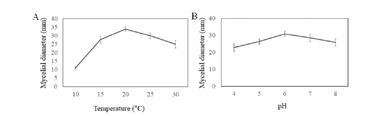
봄마름병 병원균의 온도에 따른 균사생장을 조사하기 위하여 PDA 배지에 3일간 배양된 병원균을 지름 0.5 mm 코르크보러로 균체가 포함되게 자른 다음 백금선을 이용하여 PDA 배지에 치상하고 10, 15, 20, 25와 30℃ 로 온도조건을 각각 달리한 항온 배양기에서 3일간 배양하여 균사의 직경을 측정하였다. 또한 봄마름병의 pH에 따른 균사생장을 조사하기 위하여 1 NHCl과 NaOH를 이용하여 PDA 배지 pH를 4, 5, 6, 7, 8로 조절하여 제조하였으며 병원균 균사를 치상후 25℃의 온도 조건에서 3일간 배양하여 균사생육을 조사하였다. 온도조건과 pH에 따른 균사생장은 5개 배지의 병원균 균총을 평균하여 조사하였다.
병반지 시료의 기중균사 형성 재현과 맹아출현 조사
병원균의 기중균사 형성 여부를 조사하기 위하여 병반지 잔디를 홀커터(∅80 cm)로 채취하여 25±1℃, 상대습도 90% 조건이 유지되는 생장상(DASOL SCIENTIFIC, Hwaseong, Korea)에서 기중균사 형성 여부를 관찰하였다. 가을철 봄마름병 병반지가 월동 후 봄철에 재현되는지를 확인하기 위하여, 2월초에 10홀과 14홀 2개 지역의 페어웨이에서 정상적으로 휴면이 이루어진 잔디와 마름병 병반지 잔디를 홀커터(∅80 cm)를 이용하여 각각 3점씩 시료를 채취하여 맹아형성 유무를 조사하였다. 채취된 시료는 25±1℃, 상대습도 70% 조건인 식물생장상(Daejeon, Korea)에서 3주간 잔디를 생육시켜 맹아출현 수를 세어 조사하였다.
결과 및 고찰
가을철에 발생한 봄마름병 병징
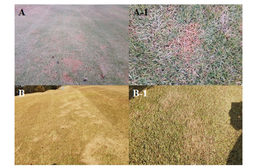
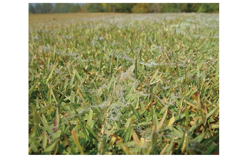
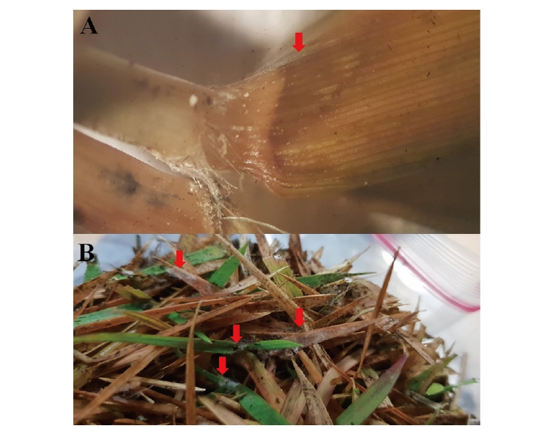
호남지역의 골프장에서 유사 봄마름병 병징이 특이하게 가을(2018년 10월 중순)에 관찰되었다. 초기병징으로 잔디의 잎이 붉은 빛으로 변하기 시작하여 10월 말경에는 30-50 cm의 원형 혹은 불규칙한 패취형태로 병반이 형성되었다(Fig. 1A and 1A-1). 이후 잔디 휴면시기인 11월 중순에 병반을 원거리에서 관찰하였을 때는 흰색이 도는 갈색이었으며(Fig. 1B) 근거리에서는 약한 붉은 색을 띄는 갈색으로 나타났다(Fig. 1B-1). 또한 이른 새벽에는 거미줄처럼 보이는 균사가 봄마름병 병반지 잔디를 덮는 경우도 관찰되었다(Fig. 2). 봄마름병은 3-4월경 그린업 되는 시기에 발병하는 것이 일반적이지만(Shim and Kim, 1995) 본 연구에서는 10월 중순에서 11월 중순까지 새롭게 병반이 형성되는 특이한 현상을 관찰하였으며 가을철 발생한 라이족토니아마름병(large patch)과 유사하여 병반의 식별이 어려웠다. 병징은 주로 엽이(leaf auricle) 부위에 나타났으며(Fig. 3A). 병반지 잔디를 홀커터(∅80 cm)로 채취하여 생장상에서 2일간 습실처리(25℃, 상대습도 90±5%)한 결과, 잔디 잎 표면에 흰색의 기중균사가 쉽게 형성되는 것이 관찰되었다(Fig. 3B).
병원균의 형태학적 특징 및 동정
봄마름병 병반부위에서 채취한 잔디시료를 염색한 후 현미경으로 관찰한 결과 잎 표면에 거미줄 모양의 병원균 균사가 관찰되었다(Fig. 4A). 분리된 봄마름병 병원균을 PDA에서 배양한 결과, 일주일간 배양된 균총은 흰색을 띄었으며 균핵을 형성하였다(Fig. 4B). 균사를 현미경으로 관찰한 결과, 균사의 격막(septum)이 뚜렷이 관찰되었고 둔각으로 분지하는 형태로 자랐으며, 균사의 직경은 6.2-7.4 μm로 관찰되었다(Fig. 4C). 병원균의 분자생물학적 동정을 위하여 ITS 영역을 분석한 결과 ITS 영역의 크기는 782 bp였으며, NCBI database에 등록된 ITS sequence중 AB198706.1, AB198704.1, AB214367.1와 95% 이상 일치하였다(Fig. 5). 병반지에서 분리된 병원균의 형태 및 유전적 특징은 Rhizoctoniacerealis AG-D와 일치하는 것으로 나타났다. 봄마름병 병원균의 온도에 따른 균사생장을 조사하기 위하여 15, 20, 25, 그리고 30℃에서 균사를 배양한 결과 20℃ 에서 34 mm로 가장 잘 자랐으며 다음으로 25℃ 와 30℃ 에서 각각 30, 25 mm 순으로 나타났다. 10℃에서는 11 mm로 가장 느린 생장이 관찰 되었다(Fig. 6A). 또한 pH 6에서 균사생장이 30 mm로 균사가 가장 잘 자라는 것으로 나타났다. Rhizoctoniacerealis AG-D가 원인이 되는 병으로는 황색마름병과 라이족토니아잎마름병이 있으며 이 병은 Rhizoctonia solani가 원인이 되는 라이족토니아마름병 보다 더 낮은 온도에서 병을 일으킨다고 보고되어있다(Kim et al., 1992; Shim and Kim, 1995).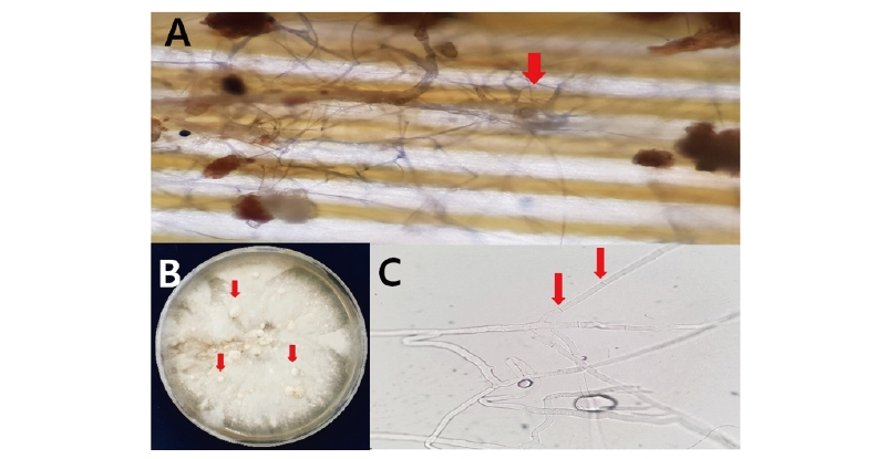
가을철 발생한 봄마름병의 발병 특징
봄마름병은 봄철에 발생하는 병으로 알려져 있지만(Tanaka, 2003) 본 연구에서는 특이하게 늦은 가을철에 발생병징이 발생하는 것을 확인하였다. 이 시기에 발생한 봄마름병의 병징(Shim et al., 1994)은 가을철 발생한 라이족토니아마름병 병징과 유사하였다. 그러나 봄마름병과 라이족토니아마름병의 차이점은 고사한 잔디의 뽑히는 현상이 다른 것으로 나타났다. 봄마름병의 경우 잔디를 당겼을 때 잘 뽑히지 않았으며 엽이 부분이 탈락되었다. 반면에 라이족토니아마름병은 잎집 부분이 잘 뽑혔다. 이러한 차이는 봄마름병과 라이족토니아마름병의 감염 부위가 달라 나타나는 현상으로 판단된다. 본 연구의 조사에서 봄마름병은 주로 엽이 부위가 썩는 현상이 나타났다(Fig. 3A and 3B). 이에 반해 라이족토니아마름병은 관부나 잎집에 병원균이 침입하여 병을 일으킨다고 보고되어있다(Shim et al., 1994).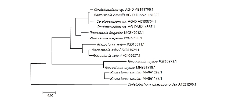
Fig. 5. Phylogenetic identification of Rhizoctonia cerealis AG-D based on internal transcribed spacer (ITS) sequences. Numerical values on branches are bootstrap values calculated as a percentage of bootstrap replications from 1000 replication. Rhizoctonia cerealis AG-D Funbio181023 represented the isolate.
가을철에 발생한 봄마름병 병반지의 맹아출현 조사
가을철에 발생한 봄마름병 병반지가 겨울철 휴면기를 거친 후 봄철에 정상적으로 맹아출현이 되는지를 확인하기 위하여 봄철에 병반지 잔디의 맹아출현을 조사한 결과, 봄마름병 병반지 시료의 잔디맹아 출현율은 건전시료 대비 약 18-28%로 낮게 나타났다(Fig. 7). 봄마름병 병반지 잔디는 6월초까지 정상적인 회복이 이루어지지 않았다. 한국잔디의 봄마름병은 가을철에 감염이 이루어져 봄철에 병징이 발현하며 방제는 감염이 이루어지는 가을철이 적기라는 보고가 있다(Hyakumachi and Hayakawa, 2008, Tanaka, 2003). 그러나 국내에서는 가을철에 봄마름병이 감염되어 병징이 발현된다는 보고는 없다. 본 연구에서는 가을철에 병징을 관찰하였고 병징으로부터 원인 병원균을 분리하여 이미 보고된 봄마름병(Shim and Kim, 1995)과 같은 병원균임을 확인하였으며 가을철에 감염 개체들이 이듬해 봄철에 맹아출현 되지 않고 병징이 발현한다는 사실도 확인하였다.
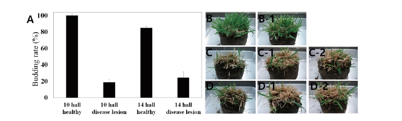
Fig. 7. Comparison of budding rate of healthy and disease region. A: Investigation of budding rate of zoysiagrass collected in golf courses fairway; B and B-1: Growth of healthy plants collected from 10 and 14 hole; C, C-1, and C-2: Growth of diseased plants collected from 10 hole; D, D-1, and D-2: Growth of diseased plants collected from 14 hole.
Authors Information
Jung Han Lee, Korea Turfgrass Research Institute, Doctor of Philosophy
Jeong Ho Kim, Korea Turfgrass Research Institute, Researcher
Chang Wook Jeon, Institute of Agriculture & Life Science, Gyeongsang National University, Ph.D. student
Gyu Yul Shim, Korea Turfgrass Research Institute, Doctor of Philosophy
Youn-Sig Kwak, Institute of Agriculture & Life Science, Gyeongsang National University, Professor